Upper Limbs
Refer to individual human bones and to the articulated human skeleton. Study the following bones and bony landmarks.
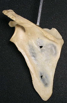
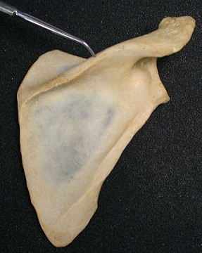
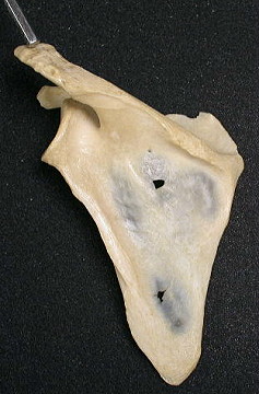
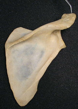
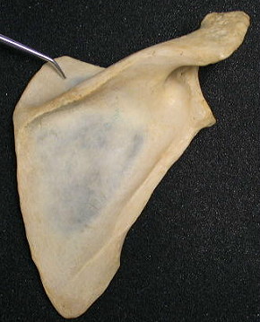
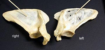
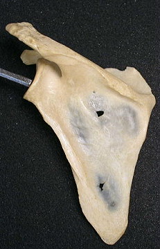
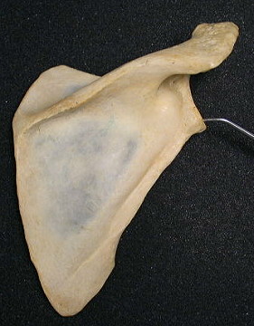
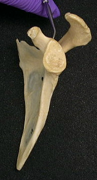
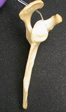
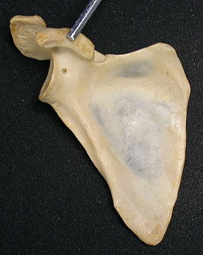
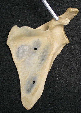
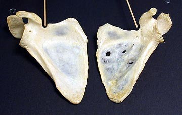
Scapula
Review the scapula. Identify the spine
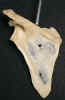
, acromion process
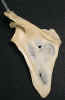
, supraspinous fossa
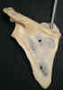
and infraspinous fossa
. Observe the glenoid cavity
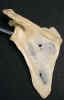
, infraglenoid tubercle
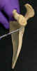
, supraglenoid tubercle
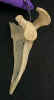
, coracoid process
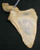
, and the suprascapular notch
. Ref. figures 1-7, 1-8 and 1-9.

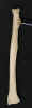
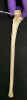
Humerus
Identify the head
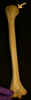
, greater tubercle
, lesser tubercle
, bicipital groove
, and the deltoid tuberosity
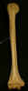
. Observe the medial epicondyle
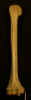
, lateral epicondyle
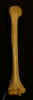
, capitulum
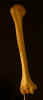
, trochlea
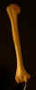
, and olecranon fossa
. Ref. figure 7-1.
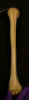
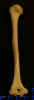
Radius
Identify the head
, neck
, radial tuberosity
, and styloid process
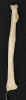
and ulnar notch
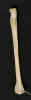
. Ref. figure 7-2.
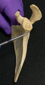
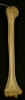
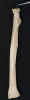
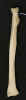
Ulna
Identify the olecranon process

, coronoid process
, radial notch
, trochlear notch
(semilunar notch) and head
. Ref. figure 7-2.
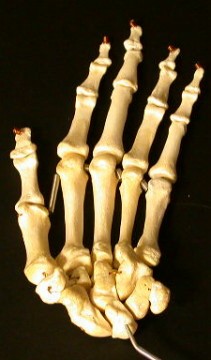
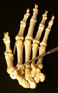
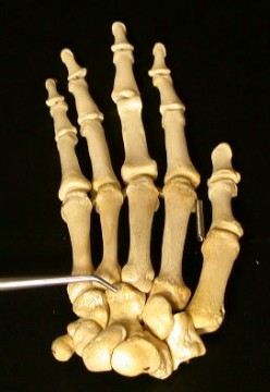
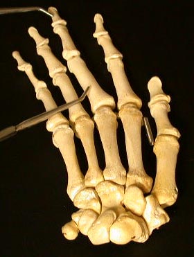
Carpus
Examine the 8 carpal bones in the articulated skeleton. Identify
in the proximal row (from lateral to medial) the scaphoid 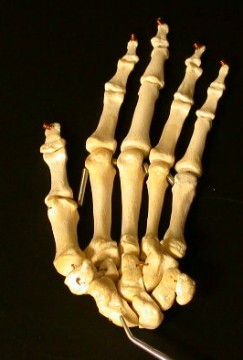
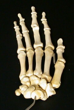 , lunate
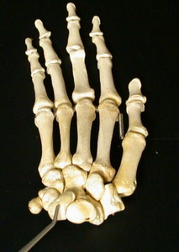 ,
triquetral
,
triquetral 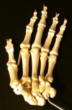
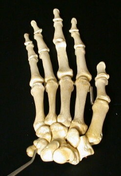 , and pisiform
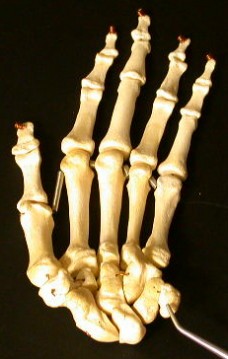
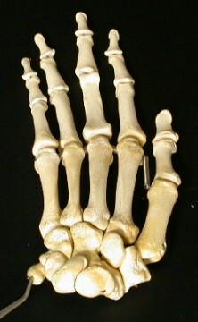 bones. In the distal row (from
lateral to medial) identify the trapezium
bones. In the distal row (from
lateral to medial) identify the trapezium 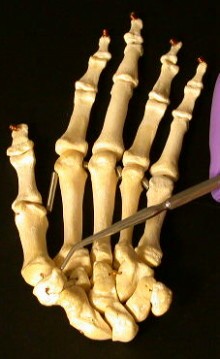
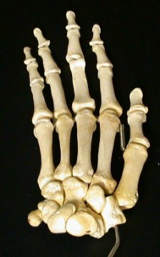 , trapezoid
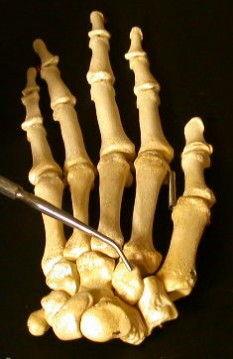 ,
capitate
,
capitate 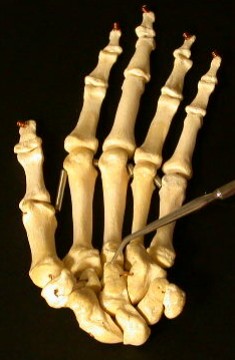
 and hamate
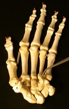
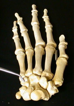 bones. Ref. figure 7-3.
Metacarpals
Observe the 5 metacarpals 
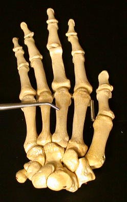 distal to the carpus.
Phalanges
Observe the phalanges 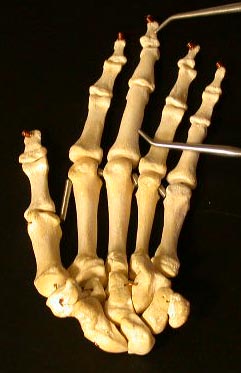
 distal to the metacarpals. The thumb
(digit 1) has only two phalanges. Digits 2 through 5 have three phalanges.
distal to the metacarpals. The thumb
(digit 1) has only two phalanges. Digits 2 through 5 have three phalanges.
Identify the above on the articulated cat skeleton.
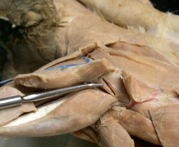
On the dorsal surface of the cat, review the deltoid and
trapezius muscles. Ref. figure 1-11. Transect the spinodeltoid
muscle and reflect it to its attachments. Identtify
the infraspinatus muscle 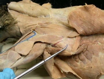
 deep to the latissimus dorsi and
spinodeltoid muscle. The infraspinatus muscle originates from the
infraspinatus fossa of the scapula and inserts on the greater tubercle of the
humerus. Observe the superficial branch of the
subscapular artery
deep to the latissimus dorsi and
spinodeltoid muscle. The infraspinatus muscle originates from the
infraspinatus fossa of the scapula and inserts on the greater tubercle of the
humerus. Observe the superficial branch of the
subscapular artery
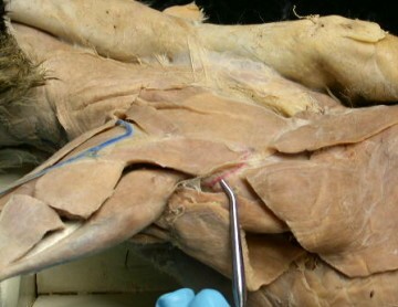
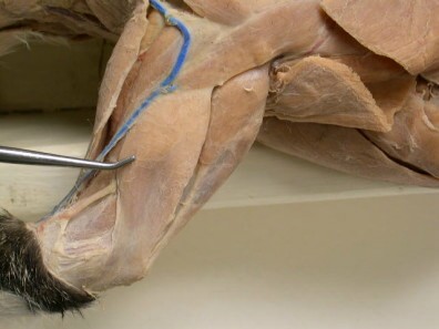
 as it emerges from between the infraspinatus and teres major
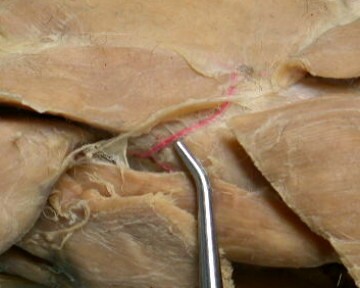
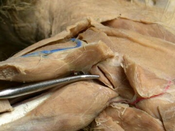
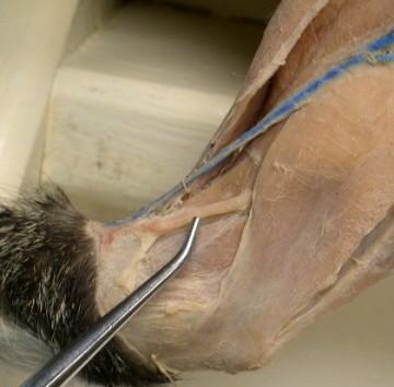
 muscles caudal to the long head of the triceps. Identify the
supraspinatus muscle
muscles caudal to the long head of the triceps. Identify the
supraspinatus muscle 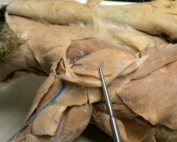
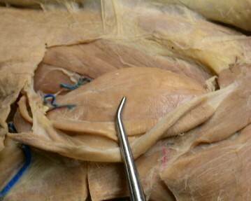 deep to the trapezius muscles. The supraspinatus muscle
originates from the supraspinatus fossa of the scapula and inserts on the
greater tubercle of the humerus. Transect across the medial
portion of the supraspinatus muscle and reflect it distally by dissecting its
origin from the supraspinatus fossa. Observe the
suprascapular
nerve
deep to the trapezius muscles. The supraspinatus muscle
originates from the supraspinatus fossa of the scapula and inserts on the
greater tubercle of the humerus. Transect across the medial
portion of the supraspinatus muscle and reflect it distally by dissecting its
origin from the supraspinatus fossa. Observe the
suprascapular
nerve 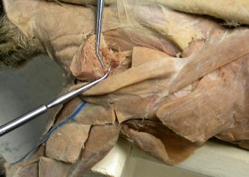
 and
suprascapular artery
as it emerges through the
suprascapular notch. Ref. figure 7-4.
and
suprascapular artery
as it emerges through the
suprascapular notch. Ref. figure 7-4.
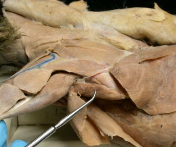
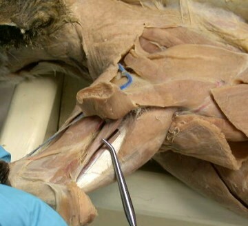
Identify the teres major muscle and the teres minor muscle. The
teres major muscle
originates from the caudal border of the
scapula ventral to the infraspinatus muscle and inserts on the proximal end of
the humerus with the latissimus dorsi muscle. The teres minor muscle
 is deep to the spinodeltoid muscle and lateral to the origin of the long head of
the triceps brachii muscle. It originates from the cranial part of the caudal
border of the scapula and passes deep to the acromiodeltoid to inserts on the
greater tubercle of the humerus. Observe the
caudal humeral
circumflex artery
is deep to the spinodeltoid muscle and lateral to the origin of the long head of
the triceps brachii muscle. It originates from the cranial part of the caudal
border of the scapula and passes deep to the acromiodeltoid to inserts on the
greater tubercle of the humerus. Observe the
caudal humeral
circumflex artery 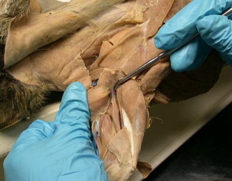
 as it emerges from between the teres minor
muscle and the long and lateral heads of the triceps brachii muscle.
as it emerges from between the teres minor
muscle and the long and lateral heads of the triceps brachii muscle.
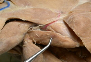
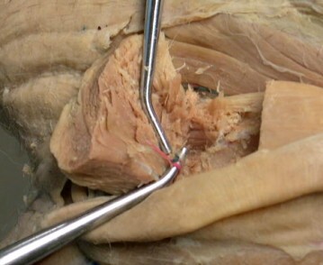
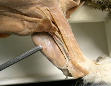
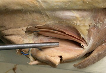
Examine the three heads of the triceps brachii muscle
on the lateral surface of the brachium. Identify the lateral
head 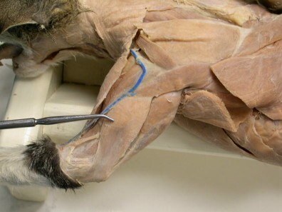
 of the triceps. It originates from the proximal end of the humerus
and inserts on the olecranon of the ulna. Isolate the lateral head
of the triceps from its origin to its insertion. Transect this
muscle and reflect it to its attachments. The medial head
of the triceps. It originates from the proximal end of the humerus
and inserts on the olecranon of the ulna. Isolate the lateral head
of the triceps from its origin to its insertion. Transect this
muscle and reflect it to its attachments. The medial head
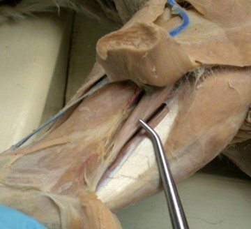 of the triceps can be seen deep to the lateral head of the triceps. It
originates from the caudomedial shaft of the humerus and inserts on the
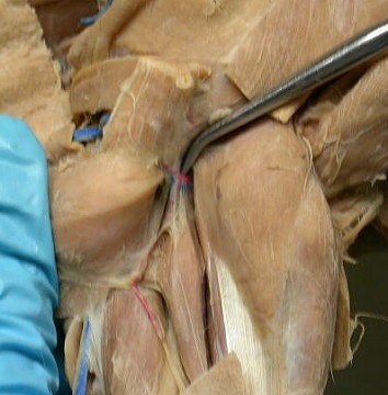
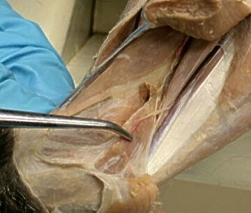
olecranon of the ulna. Observe the
radial nerve
of the triceps can be seen deep to the lateral head of the triceps. It
originates from the caudomedial shaft of the humerus and inserts on the
olecranon of the ulna. Observe the
radial nerve 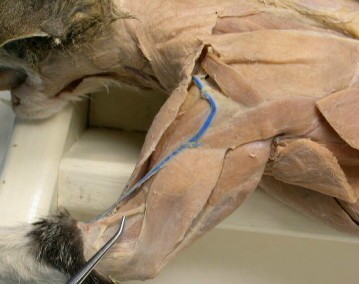
 as
it emerges from the medial head of the triceps, and courses distally, and the
caudal
humeral circumflex artery
as it passes along the medial
head of the triceps. Identify the long head
as
it emerges from the medial head of the triceps, and courses distally, and the
caudal
humeral circumflex artery
as it passes along the medial
head of the triceps. Identify the long head 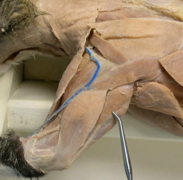
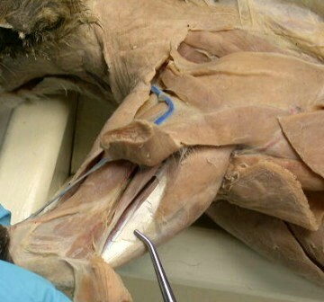
 of the
triceps. It is the largest head of the triceps and lies along the caudal surface
of the humerus. It originates from the caudal border of the scapula caudal to
the glenoid cavity and inserts on the olecranon process of the ulna.
of the
triceps. It is the largest head of the triceps and lies along the caudal surface
of the humerus. It originates from the caudal border of the scapula caudal to
the glenoid cavity and inserts on the olecranon process of the ulna.
Identify the anconeus muscle 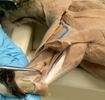
 deep to the tendon
of the insertion of the lateral head of the triceps muscle. It originates from
the lateral epicondyle and the distal lateral surface of the humerus and inserts
on the olecranon of the ulna. Identify the brachialis muscle
deep to the tendon
of the insertion of the lateral head of the triceps muscle. It originates from
the lateral epicondyle and the distal lateral surface of the humerus and inserts
on the olecranon of the ulna. Identify the brachialis muscle
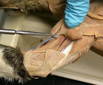
 on the lateral surface of the humerus. It originates from the craniolateral
surface of the shaft of the humerus and inserts on the proximal end of the ulna.
Its distal end is covered by the brachioradialis muscle.
on the lateral surface of the humerus. It originates from the craniolateral
surface of the shaft of the humerus and inserts on the proximal end of the ulna.
Its distal end is covered by the brachioradialis muscle.
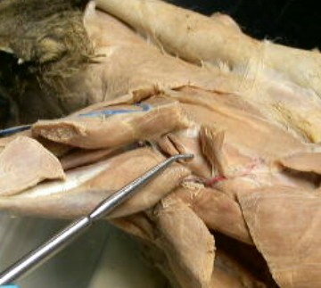
On the ventral surface, Examine the medial aspect of the
scapula. Identify the subscapularis muscle 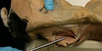
 that
originates from almost the entire subscapular fossa and inserts on the medial
aspect of the lesser tubercle of the humerus. Isolate the teres
major muscle from the subscapularis muscle. Ref. figure 7-5.
that
originates from almost the entire subscapular fossa and inserts on the medial
aspect of the lesser tubercle of the humerus. Isolate the teres
major muscle from the subscapularis muscle. Ref. figure 7-5.