We wish to acknowledge Dr. Louis Zanella's generosity in giving us permission to
include material from his dissector in this web site. It is a wonderful
addition to this study tool and we are grateful to him.
A Regional Dissector
of the Cat, Louis J.
Zanella, Ed.D.,
1996
Chapter
2: The
Thoracic Cavity
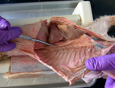
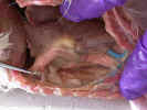
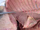
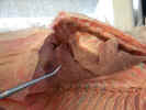
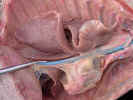
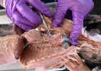
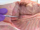
Using
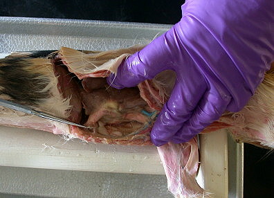
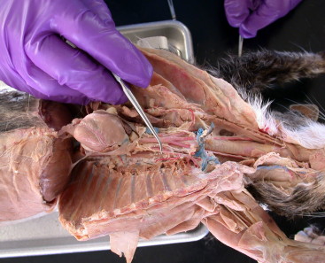
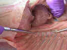
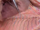
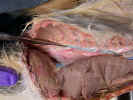
scissors open the thoracic cavity by cutting through the muscles and costal
cartilage about one centimeter right of the midsternal line. Extend this cut the
entire length of the sternum, from the first rib to the diaphragm. Spread the
incision and examine the muscular diaphragm 
 at the caudal margin of the
cavity. Make a right lateral incision cranial to and following the attachment of
the diaphragm to the ribs. With a pair of bone scissors cut the first ten ribs
near their point of attachment to the vertebrae. Do this from within the body
cavity.
at the caudal margin of the
cavity. Make a right lateral incision cranial to and following the attachment of
the diaphragm to the ribs. With a pair of bone scissors cut the first ten ribs
near their point of attachment to the vertebrae. Do this from within the body
cavity.
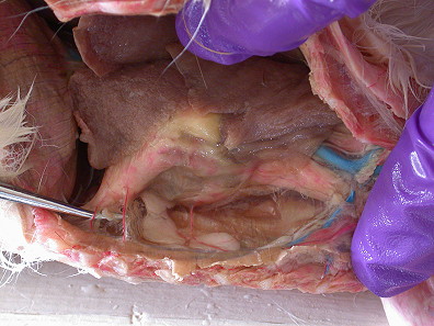
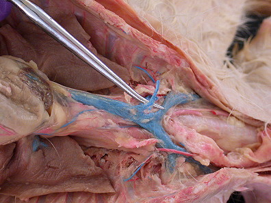
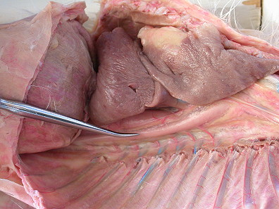
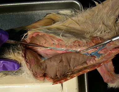
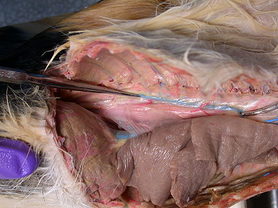
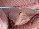
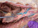
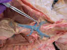
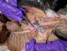
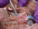
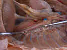
Observe
the mediastinal pleura 
 , a thin serous membrane attached to the sternum
ventrally. It extends from the dome of the neck cranially to the diaphragm and
forms the medial wall of each pleural cavity. Observe the two internal thoracic
arteries (mammary)
, a thin serous membrane attached to the sternum
ventrally. It extends from the dome of the neck cranially to the diaphragm and
forms the medial wall of each pleural cavity. Observe the two internal thoracic
arteries (mammary) 
 and the internal thoracic vein
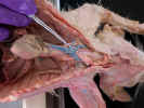
 passing through the
mediastinal pleura to the ventral thoracic wall. Clean the internal thoracic
arteries and vein and cut them at the ventral thoracic wall leaving long ends
for later study. Destroy the ventral attachment of the mediastinal pleura with
your probe.
passing through the
mediastinal pleura to the ventral thoracic wall. Clean the internal thoracic
arteries and vein and cut them at the ventral thoracic wall leaving long ends
for later study. Destroy the ventral attachment of the mediastinal pleura with
your probe.
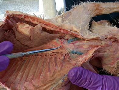
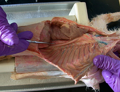
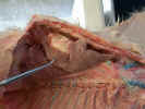
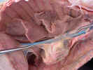
Make
a left lateral incision cranial to the diaphragm and cut the first ten ribs as
you did on the right. Spread both thoracic walls dorsally. Examine the various
parts of the pleura. The pulmonary pleura cover the surface of the lungs and is
continuous with the mediastinal pleura. The parietal pleura lines the walls of
the pleural cavity and is continuous with the diaphragmatic pleura which covers
the diaphragm. The pericardial pleura (pericardium) 
 covers the heart. The pulmonary ligament
is a pleural fold which attaches the lobes of the lungs to the medial wall of
the pleural cavity.
covers the heart. The pulmonary ligament
is a pleural fold which attaches the lobes of the lungs to the medial wall of
the pleural cavity.
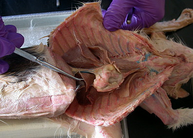
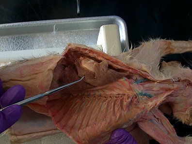
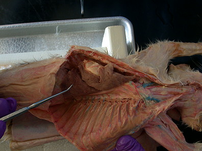
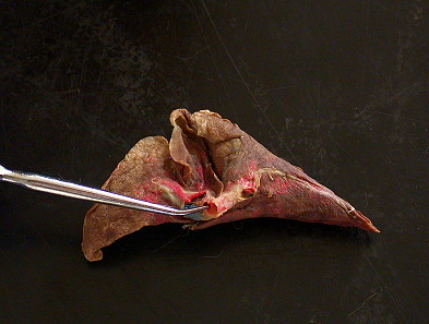
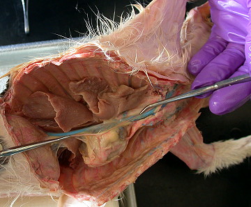
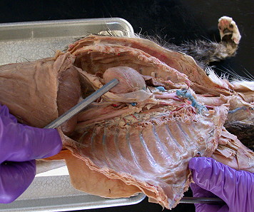
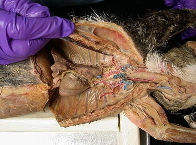
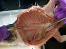
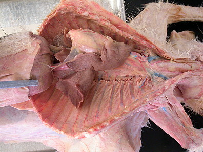
Identify
the heart, lungs, thymus and diaphragm. Examine the two lungs. The
right lung
has four lobes: cranial lobe 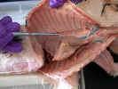
 ,
middle lobe
,
middle lobe 
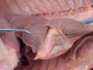 ,
caudal lobe
,
caudal lobe 
 and accessory lobe
 . The
left lung has three lobes: cranial lobe
. The
left lung has three lobes: cranial lobe 
 ,
middle lobe
,
middle lobe 
 ,
and caudal lobe
,
and caudal lobe 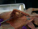
 . It does not have an accessory lobe. The apex of each lung lies 1 cm
cranial to the first rib. Between the lungs and dorsal to the heart, the trachea
. It does not have an accessory lobe. The apex of each lung lies 1 cm
cranial to the first rib. Between the lungs and dorsal to the heart, the trachea
 bifurcates into primary bronchi
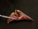
 which branch into secondary bronchi which go to
each lobe.
which branch into secondary bronchi which go to
each lobe.
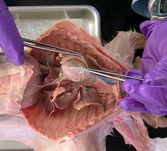
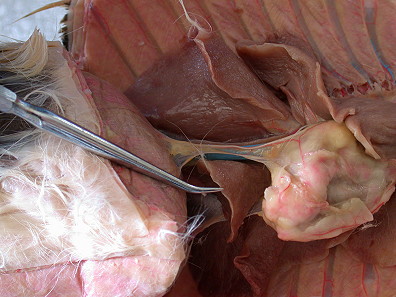
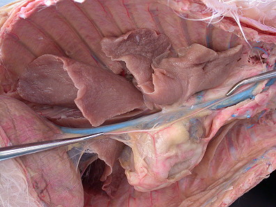
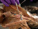
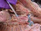
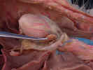
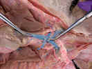
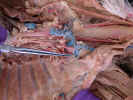
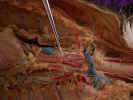
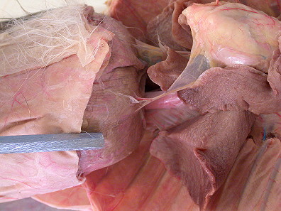
Tease
away the pulmonary ligament on the ventral surface of the root of the lung and
examine the root of the lung and observe the relationship of the bronchus,
pulmonary artery and pulmonary vein.
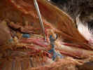
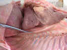
Remove
the left lung by holding all three lobes and pulling the lung laterally,
stretching the root of the lung. With scissors transect the root close to the
lung. Examine again the bronchi, the blue pulmonary
artery, and the red
pulmonary vein at the mediastinal pleura.
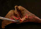
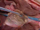
Examine the isolated lung. The primary bronchus was left
behind in the mediastinum. Identify the secondary bronchi and verify that they
contain cartilaginous rings. Hold a secondary bronchus with forceps and scrape
away lung tissue following the bronchus and its branches through the lobe.
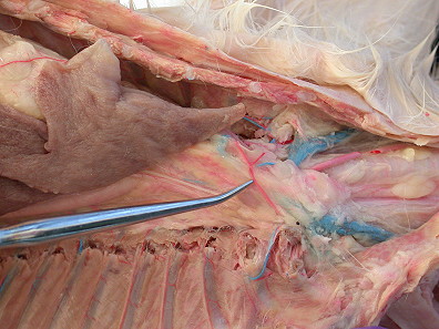
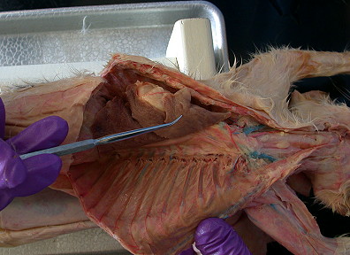
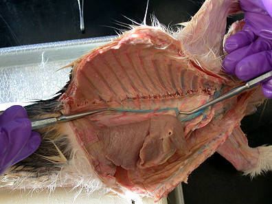
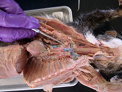
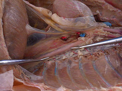
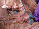
The
mediastinum is the cavity between the two pulmonary cavities. It is bound by the
cranial aperture of the thorax cranially, the diaphragm caudally, the sternum
ventrally, the bodies of the thoracic vertebrae dorsally and the lungs
laterally. This region is subdivided into a cranial, ventral, dorsal and middle
mediastinum.
The cranial mediastinum lies above an imaginary plane at
the level of the sternal angle. The remaining portion caudal to the cranial
mediastinum is divided into three parts. The ventral mediastinum is found
between the sternum and the pericardium. The middle mediastinum contains the
pericardium with the heart and the roots of the great vessels. The dorsal
mediastinum is the region dorsal to the pericardium and ventral to the thoracic
vertebrae.
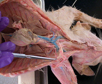
Ventral
Mediastinum
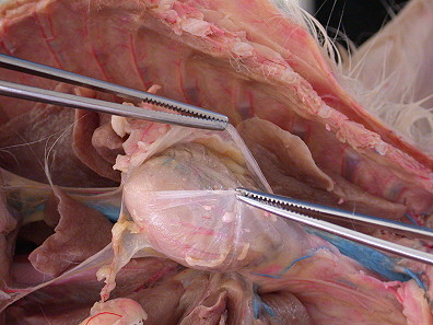
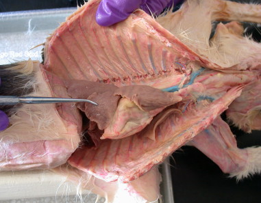
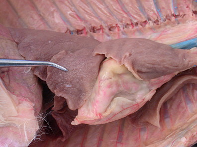
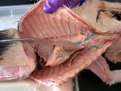
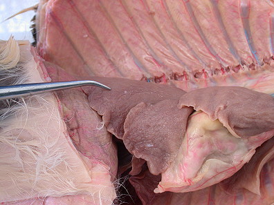
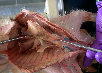
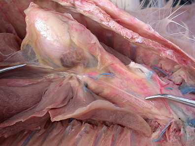
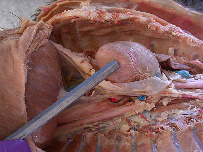
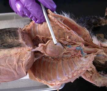
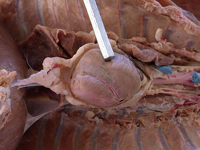
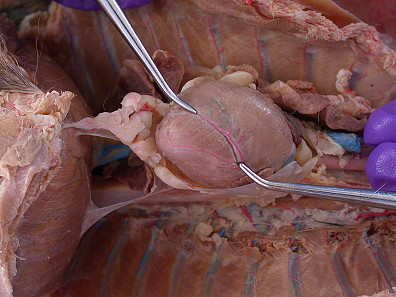
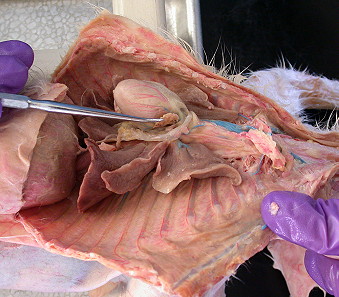
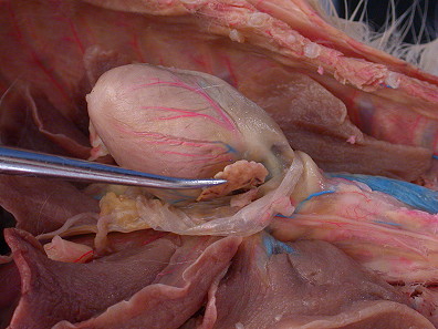
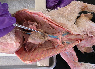
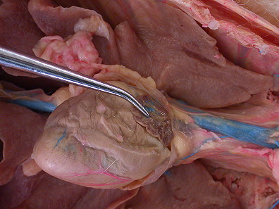
Observe the thymus gland 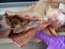
 located in the ventral and
cranial mediastinum, ventral and cranial to the heart. It is large in young
animals and decreases in size as they mature.
located in the ventral and
cranial mediastinum, ventral and cranial to the heart. It is large in young
animals and decreases in size as they mature.
Remove
the thymus being careful not to destroy the internal thoracic vessels passing
through it.
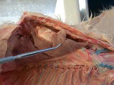
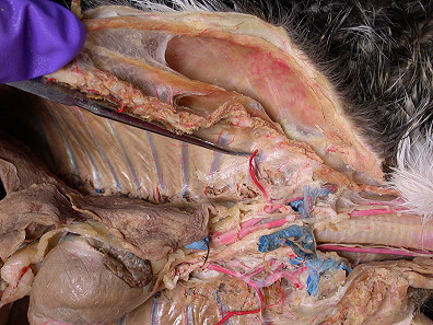
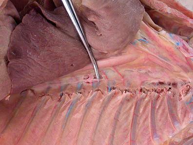
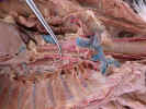
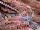
Middle
Mediastinum
The
middle mediastinum contains the pericardium, phrenic
nerves, the heart and the
roots of the great vessels. Identify the phrenic nerves


 as they pass ventral to
the root of the lungs on the side of the pericardium. Trace the left phrenic
nerve
as they pass ventral to
the root of the lungs on the side of the pericardium. Trace the left phrenic
nerve 
 caudally through the mediastinal pleura to the diaphragm. Trace the
right
phrenic nerve
caudally along the right side of the caudal vena cava
caudally through the mediastinal pleura to the diaphragm. Trace the
right
phrenic nerve
caudally along the right side of the caudal vena cava

 to the
diaphragm. Tease these nerves from the pericardium and trace them cranial to the
cranial mediastinum.
to the
diaphragm. Tease these nerves from the pericardium and trace them cranial to the
cranial mediastinum.
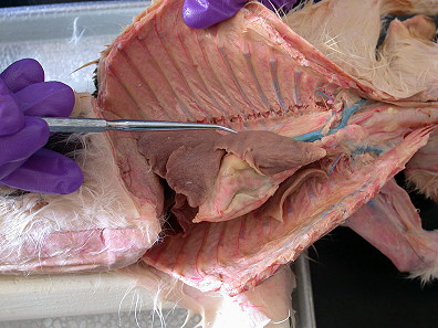
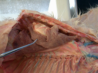
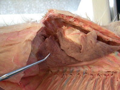
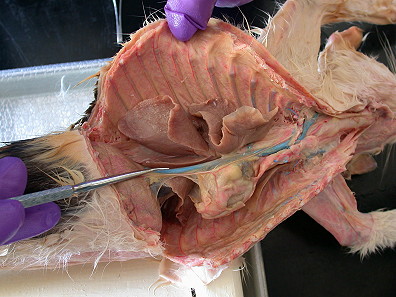
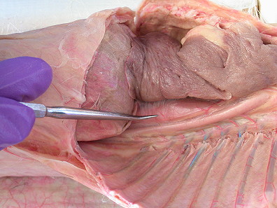
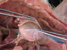
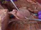
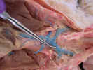
Clean
the fat from the pericardium and make a midventral incision in the pericardium
from the pointed caudal apex to the cranial base. Peel the pericardium back and
observe the shiny inner serous layer and the outer dull fibrous pericardium.
Examine
the ventral surface of the heart in situ. Observe the left ventricle 
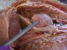 which forms
the left border and the apex of the heart and the right ventricle
which forms
the left border and the apex of the heart and the right ventricle 
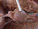 which forms
the lower border of the heart. The ventricles form the caudal two thirds of the
heart. The ventricles are separated by a shallow diagonal groove the
interventricular sulcus
which forms
the lower border of the heart. The ventricles form the caudal two thirds of the
heart. The ventricles are separated by a shallow diagonal groove the
interventricular sulcus 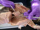
 which
contains a coronary artery and
vein. Examine the left atrium
which
contains a coronary artery and
vein. Examine the left atrium 
 at the cranial end of the left border of the heart
and the right atrium
at the cranial end of the left border of the heart
and the right atrium 
 which forms the right border. The atria are separated from
the ventricles by a groove, the coronary sulcus (this will be seen in
the heart lab). The right and left atrium are
separated from each other by the great vessels leaving the cranial end of the
ventricles. The portion of the atrium lateral to the great vessels is referred
to as the auricle.
which forms the right border. The atria are separated from
the ventricles by a groove, the coronary sulcus (this will be seen in
the heart lab). The right and left atrium are
separated from each other by the great vessels leaving the cranial end of the
ventricles. The portion of the atrium lateral to the great vessels is referred
to as the auricle.
Scrape the
fat and
remove the pericardium from the great vessels as they leave the ventricles.
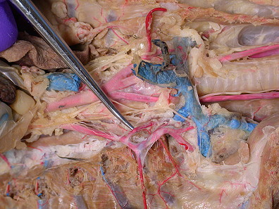
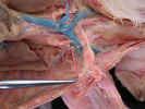
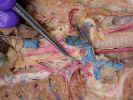
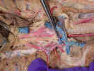
Examine the
pulmonary trunk 
 as it arises from the right
ventricle and passes cranially between the two atria. Trace it to the
bifurcation which branches to the left and right pulmonary arteries which pass
to the left and right lungs. Identify the aortic arch as it arises from the left
ventricle and passes dorsal to the pulmonary trunk.
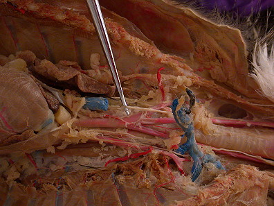
Find the ligamentum
arteriosum which connects the aorta to the pulmonary trunk at the bifurcation.
Identify the pulmonary veins caudal to the
pulmonary arteries as they leave the
lungs and enter the left atrium.
as it arises from the right
ventricle and passes cranially between the two atria. Trace it to the
bifurcation which branches to the left and right pulmonary arteries which pass
to the left and right lungs. Identify the aortic arch as it arises from the left
ventricle and passes dorsal to the pulmonary trunk.
Find the ligamentum
arteriosum which connects the aorta to the pulmonary trunk at the bifurcation.
Identify the pulmonary veins caudal to the
pulmonary arteries as they leave the
lungs and enter the left atrium.
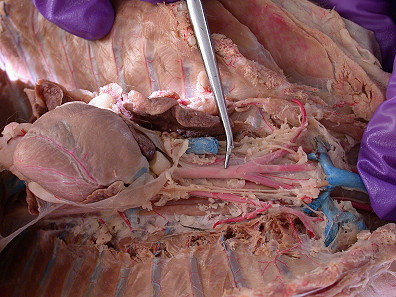
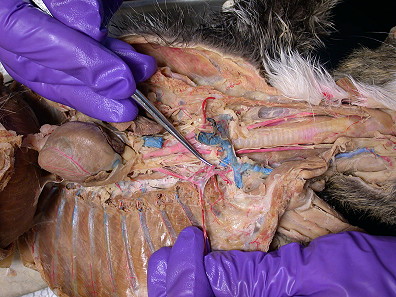
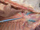
Move
the heart to the left and observe the postcava
ascending
from the diaphragm and the precava 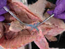
 on the right side of the neck entering the
right atrium. You will examine a sheep heart at a later date.
on the right side of the neck entering the
right atrium. You will examine a sheep heart at a later date.
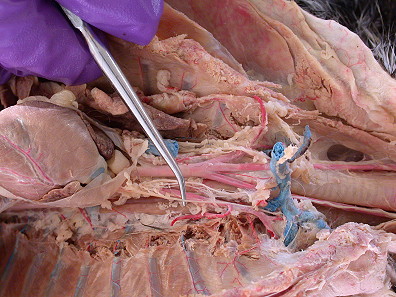
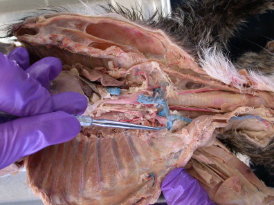
Cranial
Mediastinum
Remember
that the cranial mediastinum is only an arbitrary subdivision cranial to the
sternal angle and the base of the heart. Many structures on their way to and
from the neck pass through this space.
Clean the
cranial vena cava and trace it cranially to the left and right
brachiocephalic veins 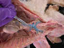

 .
Identify the azygos
.
Identify the azygos
 and internal thoracic vein
.
Cut the cranial vena cava
just cranial to the entrance of the azygos vein and
reflect the great vein
cranially to expose the aortic arch and the great arteries arising from it.
and internal thoracic vein
.
Cut the cranial vena cava
just cranial to the entrance of the azygos vein and
reflect the great vein
cranially to expose the aortic arch and the great arteries arising from it.
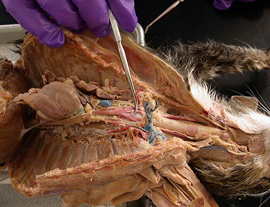
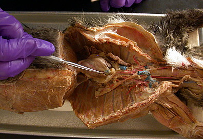
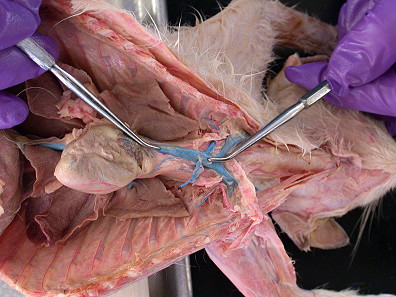
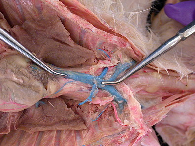
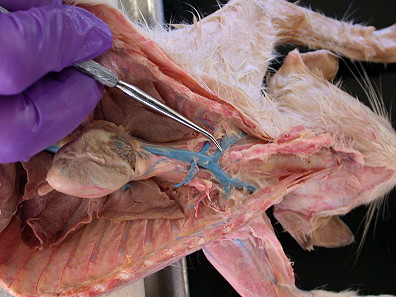
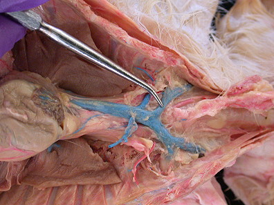
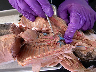
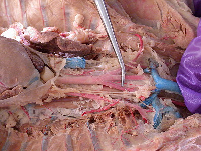
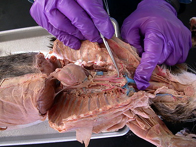
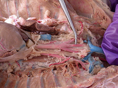
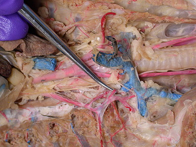
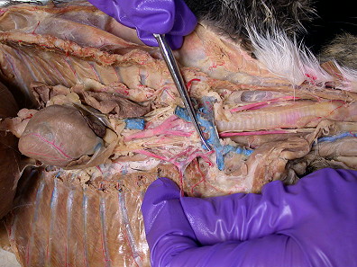
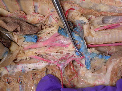
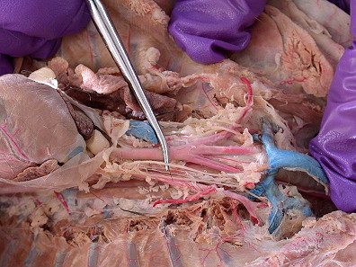
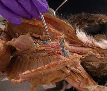
Clean and
trace the aortic arch and its branches. Trace the
aortic arch as it turns to the left and arches over the left bronchus, turns
caudally and passes dorsal to the root of the left lung. Identify the
brachiocephalic artery 
 as it arises from the aortic arch and the smaller
left
subclavian artery
as it arises from the aortic arch and the smaller
left
subclavian artery 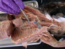
 to the left of the brachiocephalic artery. Trace the
brachiocephalic artery cranially to where it gives rise to the left
common carotid artery
to the left of the brachiocephalic artery. Trace the
brachiocephalic artery cranially to where it gives rise to the left
common carotid artery 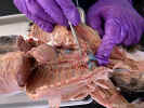
 and then the right common carotid artery
 . It
then continues as the right subclavian artery
. It
then continues as the right subclavian artery

 with the subclavian vein
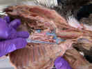
 .
.
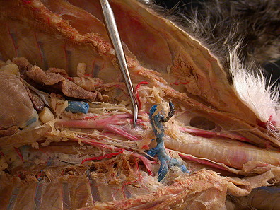
Trace the
subclavian arteries to the level of the first
rib. Identify the internal thoracic artery


 previously seen on the ventral
thoracic wall. Clean and trace the branches of the
subclavian artery distal to
the internal thoracic artery. Identify the
vertebral artery
previously seen on the ventral
thoracic wall. Clean and trace the branches of the
subclavian artery distal to
the internal thoracic artery. Identify the
vertebral artery 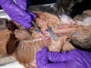
 as it arises from
the dorsal surface of the subclavian artery and passes craniodorsally to enter
the transverse foramen of the sixth cervical vertebrae. The costocervical artery
as it arises from
the dorsal surface of the subclavian artery and passes craniodorsally to enter
the transverse foramen of the sixth cervical vertebrae. The costocervical artery
 arises from the dorsal surface of the subclavian artery distal to the
vertebral artery. It sends branches to the deep muscles of the back, neck and ribs. The
thyrocervical artery
arises from the dorsal surface of the subclavian artery distal to the
vertebral artery. It sends branches to the deep muscles of the back, neck and ribs. The
thyrocervical artery 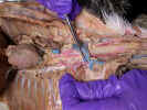
 arises from the subclavian artery distal to the
costocervical artery and passes laterally and cranially supplying muscles of the
neck and shoulders.
arises from the subclavian artery distal to the
costocervical artery and passes laterally and cranially supplying muscles of the
neck and shoulders.
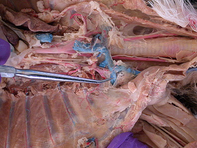
Trace
the left phrenic nerve
cranially as it crosses the aortic arch and enters the
thoracic cavity near the subclavian artery. Trace the
right phrenic nerve
cranially along the right side of the trachea close against the precava to where
it enters the thoracic cavity.
Identify
the vagus nerves with the common carotid arteries and the
internal jugular veins. Trace the left vagus nerve
 caudally as it crosses the lateral surface of
the aortic arch. It continues dorsal to the root of the left lung where it gives
rise to cardiac branches. Trace the
right vagus nerve
caudally as it crosses the lateral surface of
the aortic arch. It continues dorsal to the root of the left lung where it gives
rise to cardiac branches. Trace the
right vagus nerve 
 caudally along the right
of the trachea and dorsal to the root of the right lung where it gives rise to
cardiac branches. Caudal to the tracheal bifurcation each nerve divides into
dorsal and ventral branches. The dorsal branches from the
left and right vagus
nerve unite to form the dorsal vagal trunk
caudally along the right
of the trachea and dorsal to the root of the right lung where it gives rise to
cardiac branches. Caudal to the tracheal bifurcation each nerve divides into
dorsal and ventral branches. The dorsal branches from the
left and right vagus
nerve unite to form the dorsal vagal trunk
 and the ventral branches form the
ventral vagal trunk which lie on the dorsal and ventral surfaces of the
esophagus respectively.
and the ventral branches form the
ventral vagal trunk which lie on the dorsal and ventral surfaces of the
esophagus respectively.
Above
the level of the aortic arch observe the trachea and the esophagus. The
esophagus lies ventral to the vertebral column and slightly to the left of the
trachea. The trachea
is immediately ventral to the vertebral columns ventral to
and to the right of the esophagus. Identify the individual tracheal rings.
Trace
the esophagus caudally through the dorsal mediastinum to the diaphragm.
Dorsal Mediastinum
Displace the heart to the right and
examine the
dorsal mediastinum, from the left side. Observe the descending
aorta 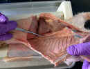
 .
Tease away the pleural ligament to the aorta and observe the
esophagus
.
Tease away the pleural ligament to the aorta and observe the
esophagus 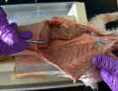 ventral to the aorta. Identify the ventral vagus trunk
behind the root of the left lung. Free and trace it caudally along the ventral surface of the esophagus.
Identify the thoracic duct
ventral to the aorta. Identify the ventral vagus trunk
behind the root of the left lung. Free and trace it caudally along the ventral surface of the esophagus.
Identify the thoracic duct
 found dorsal and to the left of the aorta. Trace the thoracic duct through the
cranial mediastinum. Tease the parietal pleura lateral to the vertebral column
and find the sympathetic trunk
found dorsal and to the left of the aorta. Trace the thoracic duct through the
cranial mediastinum. Tease the parietal pleura lateral to the vertebral column
and find the sympathetic trunk
 which lies behind the pleura. Trace it
from the diaphragm cranially through the cranial mediastinum.
which lies behind the pleura. Trace it
from the diaphragm cranially through the cranial mediastinum.
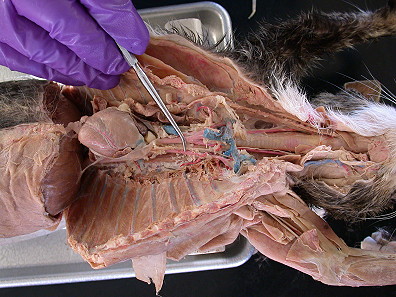
Pull the right lung ventrally and
examine the right
sympathetic trunk. Identify the dorsal vagus trunk dorsal
to the root of the lung and trace it to the dorsal aspect of the
esophagus
.
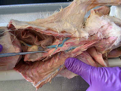
Identify the
azygos vein 
 close against the vertebral column. Trace the azygos cranial to
the root of the right lung where it crosses over and empties into the cranial
vena cava.
close against the vertebral column. Trace the azygos cranial to
the root of the right lung where it crosses over and empties into the cranial
vena cava.
 at the caudal margin of the
cavity. Make a right lateral incision cranial to and following the attachment of
the diaphragm to the ribs. With a pair of bone scissors cut the first ten ribs
near their point of attachment to the vertebrae. Do this from within the body
cavity.
at the caudal margin of the
cavity. Make a right lateral incision cranial to and following the attachment of
the diaphragm to the ribs. With a pair of bone scissors cut the first ten ribs
near their point of attachment to the vertebrae. Do this from within the body
cavity.